- What is physical activity?
- Energy expenditure
- Movement
- Posture
- Volume, intensity, duration, frequency
- Physical behaviour type
- Contextual information: Domain, spatial settings and social contexts
- Sedentary behaviour
- Physical activity guidelines
- Physical activity variation
- Inventory and taxonomy of pattern metrics
- Introduction to objective methods
- Pedometers
- Accelerometers
- Heart rate monitors
- Combined heart rate and motion sensors
- Direct observation
- Doubly labelled water
- GPS and other GNSS receivers
- Multi sensor monitors
- Harmonisation of physical activity data
- Case study: Physical activity during pregnancy and anthropometry of the offspring
- Comparison of three harmonisation methods using validation data
- Network harmonisation of physical activity data using validation data
- Physical Activity Assessment Video Resources
- Getting participants started with the Axivity Monitor
- Wearing the Axivity Monitor
- Getting participants started with the ActiGraph Monitor
- Getting participants started with the Actiheart Monitor
- Wearing the ActiGraph monitor
- Step Test Procedure
Accelerometers
Accelerometers are instruments which measure acceleration, the change in velocity of an object over time (SI unit: m.s-2). Acceleration is directly proportional to the force acting on the object to move it (as is the mass of the object).
Physical activity can be regarded as the change of position of body segments resulting from skeletal muscle contractions. A measure of acceleration of body segments can therefore be used to infer intensity of physical activity over time, allowing derivation of activity dimensions such as duration, frequency and overall volume. If acceleration and mass (including any external loading) of all major body segments are measured, whole-body energy expenditure due to physical activity can be estimated using the work-energy theorem. In practice, we seldom measure acceleration of the whole body or have any knowledge of external loading; this imperfect knowledge is therefore a source of error when inferring energy expenditure from accelerometry data. Like most other objective methods, accelerometers do not provide contextual information. Methods to identify activity type from raw accelerometry have been proposed (Janidarmian et al., 2016; Preece et al., 2009; Willetts et al., 2018).
Table P.3.5 Physical activity dimensions which can be assessed by accelerometer.
| Dimension | Possible to assess? |
|---|---|
| Duration | ✔ |
| Intensity | ✔ |
| Frequency | ✔ |
| Volume | ✔ |
| Physical activity energy expenditure | ✔ |
| Type | ✔ |
| Timing of bouts of activity (i.e. pattern of activity) | ✔ |
| Domain | |
| Contextual information (e.g. location) | |
| Posture | ✔ |
| Sedentary behaviour | ✔ |
Accelerometer types
Technological advances have resulted in instruments that can measure acceleration accurately, over extended time periods (a week or longer), and that are sufficiently compact and discrete for people to wear. All accelerometers have two essential parts: 1) a transducer or sensor which senses acceleration; and 2) a data acquisition system which processes and stores the data. The options for the sensor component fall into two basic types:
- Piezo-electric sensors
- Natural phenomenon measured is voltage
- Low power consumption
- Must be calibrated before use
- Sensitive to acceleration in dynamic, but not in static, situations. Gravitational acceleration is therefore only transiently present in the data when the monitor is rotated relative to gravity, thus not possible to assess posture
- Digitisation and filtering to reflect relevant acceleration component
- Output may be stored as raw waveform in S.I. units but for older instruments data are often stored as proprietary “counts” in reduced time resolution
- Seismic or inertial sensors (aka MEMS)
- Natural phenomenon measured is capacitance
- Sensitive to acceleration in static and dynamic situations
- Digitisation to reflect acceleration (can be calibrated on-the-fly using gravity in still segments)
- Output usually as raw waveform in S.I. units but can also be reduced to proprietary “counts”
Accelerometer models
Accelerometers come in various models and specifications, and from many different manufacturers (brands) (Welk, 2000). It is important to emphasise that validity is tied to the overall method, rather than solely to the accelerometer make or brand. The properties of the captured data depends on number of acceleration axes, piezo-electric or MEMS-based, resolution and dynamic range, anatomical attachment site, and degree of filtering / data processing (if any) before data storage. The method includes all these components but also subsequent data processing decisions of feature extraction and inference.
It is not possible to recommend one device over another without knowing the specific aims of a particular study; however, the more information that is captured, the greater the options are for developing or applying more sophisticated methods to estimate specific variables of interest. Links to specific instruments are provided in the instrument library. The following should be considered when choosing between accelerometers:
- Raw or count-based accelerometry (raw is device agnostic)
- Evidence of technical reliability
- Robustness for use in field settings
- Evidence of validity for proposed inference method
- Ability to process the data to extract target variables
- Cost and budget
- Burden upon participants, e.g. size, wear location
- Data storage capacity
- Number of days sampling required
Axes
Accelerometers can measure acceleration in one, two or three directions, correspondingly denoted uni-axial, bi-axial, and triaxial accelerometers (Chen et al., 2012). Naturally, it is preferable to measure all three dimensions of the physical world (otherwise the device is “blind” to movement in one or two directions), and the majority of contemporary instruments are capable of this. Inference based on uni- and bi-axial acceleration implicitly relies on pre-dominance of importance of one or two of the axes, or cross-axes correlations to capture the complete physical movement of the body segment to which the instrument is attached. Tri-axial accelerometers are therefore more sensitive to certain types of activity where movements are more variable in three-dimensional space, such as climbing, jumping or spontaneous play (Ott et al., 2000).
Sampling frequency and data storage
Modern accelerometers have sufficient storage capacity to store acceleration signals at frequencies sufficiently high to reproduce the acceleration waveform over multiple days (thus, sometimes referred to as waveform accelerometers). For example, the instrument used in the UK Biobank study is capable of storing tri-axial acceleration at 100 Hz for 14 days (Axivity, 2013). Battery life and storage capacity permitting, recording sampling frequency should be as high as possible to enable a wider range of methods.
Due to memory and battery limitations, some older accelerometer instruments initially sample acceleration at frequencies between 10-32 Hz before conducting on-board data processing and feature extraction, summarising signals ‘on-the-fly’ as counts stored at a user-defined epoch. As above, shorter epoch durations are desirable to optimise the resolution of the captured information as data can be down-sampled if required; however in some instruments only minute-by-minute resolution is available which still provides over 10,000 measurements in a week. Regardless of the resolution of raw stored data, a separate decision can be made on the analysis epoch duration; the lower limit of this is constrained by the resolution of the raw data but it can otherwise be tailored to the research question. A common analysis epoch for raw waveform acceleration data is 5 seconds, as for example used for the summarised movement intensity data in the UK Biobank data showcase (Doherty et al. 2017) or WHO surveys (Westgate et al., 2019).
Monitor placement
The body segment that the accelerometer is attached to is a strong determinant of what information is captured and recorded. The interpretation of the resulting signal has to take the anatomical placement into account, because the biomechanical profile of different activity types determines the relationship between the acceleration of one body segment and the acceleration of the rest of the body segments.
The most appropriate position of an accelerometer depends on the study question and feasibility considerations. The hip or lower back allows for tracking the movement of the largest and most central part of the body, the trunk. A seismic accelerometer attached to the thigh may be used to estimate posture from its orientation with respect to gravity. The wrist is generally considered to be the most acceptable wear location to participants during free-living monitoring, and is therefore increasingly used in surveillance systems where representativeness of the sample is a particular concern.
During some activities, the acceleration of one body segment may not be representative of other body segments or the body as a whole; one such example of this is upper and central body movement whilst cycling, where measurement of acceleration at the wrist or trunk would likely result in under-estimating energy expenditure.
Number of monitors
As indicated above, the data that can be captured by a single accelerometer only yields incomplete information about an individual’s physical activity across a range of activity types. More complete information can be collected by measuring acceleration at two or more body locations simultaneously, which may enhance inferences about physical activity. However, it is only recently that accelerometer hardware and price have reached the point where multi-monitor measurement might be considered a feasible option for population research, so it has not yet been implemented in any large scale studies; accordingly, development of the methodology for interpreting multiple acceleration signals in a complementary manner has attracted considerably less attention but there are examples (Staudenmayer et al., 2009, Straczkiewicz et al., 2016, White et al., 2019).
Using labelled data collected in a laboratory, classification (e.g. machine learning) models utilising raw acceleration signals recorded at multiple body sites are better able to discriminate between different activity types (Bao & Intille, 2004, Preece et al., 2009a, Preece et al., 2009b). Another study showed that models based on acceleration measured at wrist and thigh locations had slightly lower error in predicting activity energy expenditure during free-living than either signal alone (White et al., 2019). Further methodological work is required to understand how best to implement and interpret different monitor configurations.
Naturally, the increase in information data capture has to be weighed against the increased burden upon the participant, which can cause issues with reactivity and reduce protocol adherence.
Duration of monitoring period
If inferences are to be made about habitual physical activity (whether this is in terms of volume, type, intensity etc.), it is necessary to consider the duration of monitoring, ie number of days of measurement required to capture a stable average estimate of habitual activity, given day-to-day within-person variation in physical activity. Minimum criteria can be applied based on the within-and between-individual variation of the variable of interest, as described in the section on physical activity variability.
Accelerometry is the most common objective method used to measure physical activity in population studies; it has been used extensively in field settings for:
- Large-scale observational cohort studies examining associations between activity exposure(s) and outcome(s)
- Surveys describing levels and temporal patterns of activity by population subgroups
- Interventions and randomised controlled trials examining treatment efficacy
- Personal monitoring of activity behaviours by the general public using commercial wearables and smartphone-based activity tracker apps
Accelerometers were initially used as an outcome measure in small studies and a criterion measure to compare with self-report data. As accelerometers have become cheaper they have increasingly been used in large studies (Wijndaele et al., 2015), including thousands of participants in the Pelotas birth cohorts (da Silva et al., 2014) and National Health and Nutrition Examination Survey (NHANES) (Troiano et al., 2014), and 100 thousand in UK Biobank (Doherty et al., 2017). In addition, commercial wearables, smartphones, and other "smart" everyday devices such as cameras and toothbrushes include accelerometers.
When raw acceleration data is provided in SI units (m.s-2), processing data and estimation of dimensions of physical activity may follow a series of user-defined steps, all of which affect the validity of the final inference.
The measured value of a raw acceleration signal contains three components:
- Acceleration as a result of human movement – the critical component when estimating physical activity
- Acceleration as a result of gravitational force - in static situations this tells us the orientation of the (triaxial) accelerometer
- Noise
In order to assess physical activity, one may wish to isolate the human movement component of the signal (van Hees et al., 2012). Interpreting this signal in a reliable way, and the methodology for doing so is still under active development. While there is no consensus on standard procedures for making such inferences, an example pipeline is summarised in Figure P.3.3, which is described in detail below.
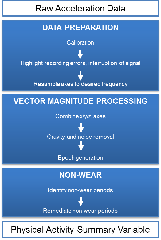
Figure P.3.3 Commonly applied processing steps for deriving physical activity summary variables from raw acceleration data. Adapted from: Doherty et al., 2017.
Data preparation
Preparatory steps involved in processing raw accelerometry data include:
- Calibration of acceleration signals to local gravity to compensate for hardware inconsistencies and provide a standardised measure of acceleration (Lukowicz et al., 2004; van Hees et al., 2014).
- Highlight recording errors, interruption of signal and instances when the sensor’s dynamic range is exceeded.
- Optional resampling of the measured signal to a chosen rate (sampling rate can fluctuate around a defined rate).
Vector magnitude processing
These steps summarise the tri-axial acceleration movement component as a single-value average over an analytical epoch duration defined by the researcher.
- Combining the x, y and z axes to calculate the vector magnitude (VM) using the formula VM = (x2 + y2 + z2)0.5, see Figure P.3.4. VM thus represents the resultant acceleration that the device is subjected to at any time point, irrespective of direction.
- Removing gravity from VM using one of several available approaches (see Figure P.3.5):
- Euclidean norm minus one (ENMO): subtract 1g (one gravitational unit) from the vector magnitude and truncate any resulting negative values to zero (van Hees et al., 2015).
- High-pass filtered VM (HPFVM): use a high-pass filter to remove low-frequency components of the signal, including gravity.
- Removing noise using a low-pass filter. Signal chances above a certain frequency are assumed not to be the result of bodily movement. This noise could be a result of external mechanical forces or the instrument itself. Human movement is typically in the range 0-15 Hz, so a low-pass filter can remove the high-frequency noise from the desired physical activity signals. To apply a low-pass filter with a cut-off of 15 Hz, the sampling frequency must be >30 Hz according to the Nyquist sampling theorem (twice the cut off).
- (Optional) Calculate the movement intensity signal in analytical epochs. The average of all activity-related resultant acceleration values within the defined time interval (e.g. each 5 seconds).
- (Optional) Summarising the movement intensity signal across the whole monitoring period for the individual, e.g. as average movement intensity (volume) or time spent at different movement intensity levels (participant-level summary variables, see below)
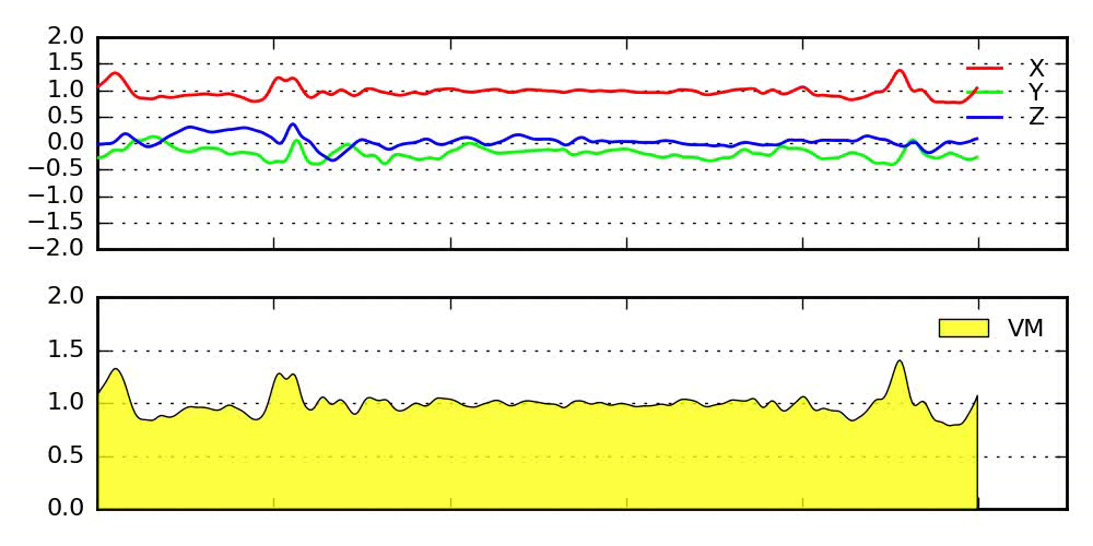
Figure P.3.4 Calculation of the vector magnitude (VM) using the Euclidean norm of the x, y and z axes.
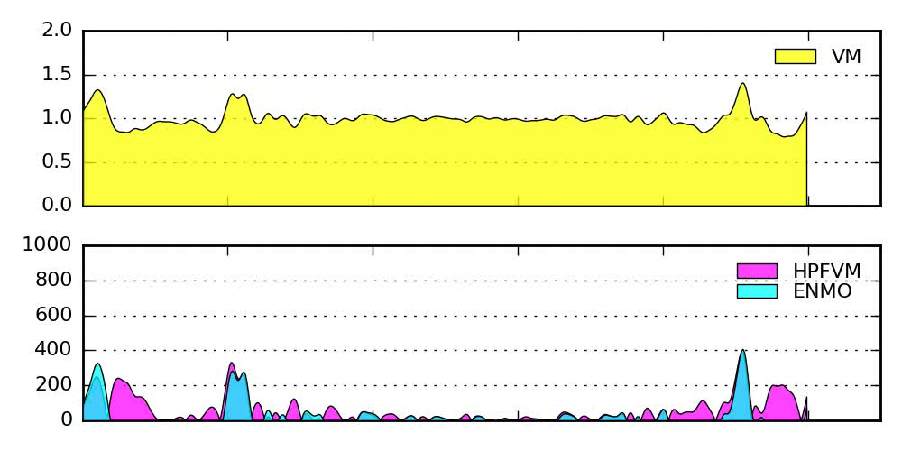
Figure P.3.5 Use of high-pass filter (HPFVM) and Euclidean norm minus one (ENMO) techniques to remove gravity component of raw acceleration signal.
Non-wear time
It is important to recognise that regardless of the wear protocol dictated by the study design, there are likely to be periods where study participants have not worn their device. These periods can be identified in a number of different ways.
- Using a device log to capture non-wear episodes. This method has higher participant burden and carries higher risk of reactivity to being observed.
- Identification of non-wear time using a user-defined algorithm, for example 10-100 consecutive minutes of stationary time (Atkin et al., 2013). For raw acceleration data, stationary episodes can be identified using the standard deviation of the acceleration of the three axes and a threshold value which accounts for the intrinsic noise level of the sensor (e.g. <13 mg = stationary) (van Hees et al., 2013). Algorithms for detecting non-wear time using count based accelerometers have also been developed (Cain et al., 2013; Choi et al., 2011; Hutto et al., 2012). The difference between true non-wear and physical inactivity (e.g. during sleep or rest) is shown by Figure P.3.6.
- As with other objective methods which involve body-worn instruments, decisions must be made regarding how non-wear time is handled once identified; it is essentially a missing data problem. Please see the dedicated section on this topic.
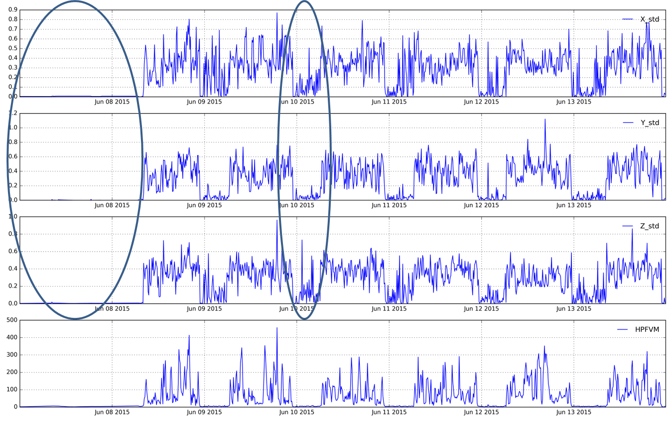
Figure P.3.6 Difference between true non-wear (left) and wrist acceleration during sleep (right). Panels are x, y, z, and high-pass filtered vector magnitude.
Brand-specific software packages are available which enable users to process data, as described in the section below. In some cases, it is also possible to export data and conduct all or some of the above steps in freely available software.
Physical activity summary variables
The steps in Figure P.3.3 result in a summary of physical activity as mean acceleration (g or mg) per user-defined epoch. For count-based accelerometers the unit will be in counts per user-defined epoch. Additional variables such as average acceleration by day and hour, or time spent at different acceleration levels at participant level (see Figure P.3.7) can be derived to investigate patterns of activity and compare individuals.
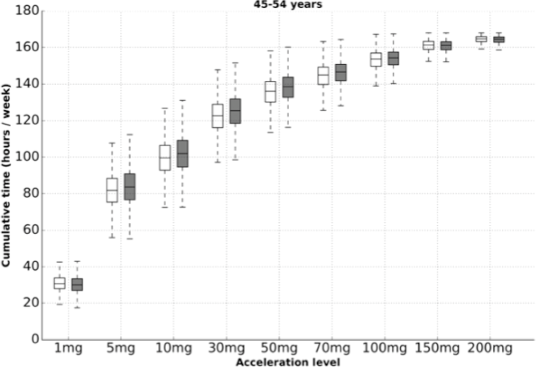
Figure P.3.7 Cumulative time spent in various movement intensities by sex for 45-54 year old women (white) and men (grey) in the UK Biobank Study. Source: Doherty et al., 2017.
Note on processing proprietary “count” accelerometer data
Older accelerometer store data in proprietary formats (i.e. counts), and several processing steps occur on-board the instrument. The details of these on-board processing steps are often known only to the manufacturer and are not decided by the user. It is important to acknowledge that these data processing ‘decisions’ made by any given monitor makes the stored information fundamentally different to the original acceleration signal. It is also important to acknowledge that some older count-based accelerometers capture movement in only one or two directions.
Further inferences
Physical activity energy expenditure
Accelerometers do not directly measure activity energy expenditure but there is a natural relationship between bodily movement and energy expenditure which can be exploited by predictive models. In practice, the characterisation of this relationship is complex because it varies by the body segment being measured and the activity type being performed.
The relationship between accelerometer recordings and energy expenditure is often studied by collecting data in a laboratory, where participants can be measured contemporaneously with a gold-standard measure of energy expenditure, such as respiratory gas analysis using facemask/mouthpiece or inside a calorimeter. In a typical study design, participants are often asked to perform a set routine of activities of varying intensity (Freedson et al., 1998; Swartz et al., 2000; van Hees et al., 2011) .
The overall relationship between activity energy expenditure and uniaxial acceleration measured at the waist during rest, walking, and jogging is fairly linear; however, deviations from linearity occur for high-intensity running (Brage et al., 2003a; Cavagna et al., 1976), for which movements are better captured with additional measurement of the antero-posterior axis of acceleration (Brandes et al., 2012). Linear relationships derived for rest and ambulation show much poorer validity in biomechanically diverse activities, such as cycling or lifting weights. Non-linear statistical models have been proposed to improve prediction equations. For example, a 2-segment regression model has been shown to improve accuracy of energy expenditure estimates for some activities, compared with simple regression (Crouter et al., 2006).
Those intending to use models derived by laboratory studies should critically evaluate the study population, and judge how appropriate it is to generalise from the activities performed in the lab setting. Laboratory studies may not reflect relationships between acceleration and energy expenditure in free living, and laboratory-derived prediction equations have been found to substantially under- or overestimate free-living energy expenditure if implemented non-critically (Corder et al., 2008; Ellis et al., 2016).
The validity of different accelerometer methods have been reviewed, demonstrating large variability in mean bias and correlation with estimates of energy expenditure using the doubly labelled water (DLW) method (Plasqui & Westerterp, 2007). Studies using DLW have shown that, in children, accelerometers explained 13% of DLW-measured PAEE variance and 31% of TEE variance. In adults, explained variance was higher, 29 and 44% for PAEE and TEE (Sardinha & Judice, 2016). Studies have also shown differences in values both within- and between-models (Brage et al., 2003b; Freedson et al., 2005; Ried-Larsen et al., 2012).
Using “cut-points” to estimate time spent in various intensity categories
A model of the relationship between acceleration magnitude and rate of energy expenditure (intensity) can be used to derive “cut-points” that correspond to a given energy expenditure value. For example, a researcher interested in quantifying time spent at or above “moderate” intensity may want to find acceleration intensity cut-point that best discriminates between < 4 METs and > 4 METs.
However, cut-points for defining different intensity levels in relative metabolic terms are somewhat arbitrary and the use of different cut-points can have a profound impact on estimates of physical activity (Freedson et al., 2005, Matthew, 2005). A researcher using accelerometry must understand the derivation of prediction equations from calibration studies and the rationale and implications of choosing a particular set of cut-points (Matthew, 2005; Rowlands et al., 2013 ). For example, published cut-points for sedentary behaviour from one accelerometer vary from 100 cpm to 1100 cpm (Atkin et al., 2013). Similarly, the range of cut points for moderate-intensity activity varies between 200 cpm to 3000 cpm.
Overall, cut-points make limited use of the wealth of detail available from raw acceleration data, often sufficient only for a crude approximation of the intended outcome (Matthews, 2005). Given the arbitrary nature of the count-based cut-offs, and the difference in unit expression across accelerometer models, reporting accelerometry data in standardised units of acceleration (m.s-2) is recommended (Brage et al., 2003a; Corder et al., 2008; Freedson et al., 2005). Extraction of signal features and patterns from raw acceleration data may improve PAEE estimation, and offers the ability to make inferences about posture from limb angles (Rowlands et al., 2015).
Posture
Using raw accelerometer data, it is possible to make inferences about an individual’s posture. When the instrument is stationary, it should measure 1g of acceleration in total over all 3 axes. The ratio of X:Y:Z indicates the direction gravity is acting on the device. From this, pitch and roll can be calculated (assuming the accelerometer X and Y axes align with a body segment's transverse and longitudinal axes, respectively):
- Pitch = arctan( Y / √(X2; + Z2) ) x (180 / Π)
- Roll = arctan( X / √(Y2 + Z2) ) x (180 / Π)
One way to think about this is to liken the accelerometer to a spirit level. For example, placing an accelerometer on the thigh will allow us to measure thigh pitch, i.e. angle with horizontal; this feature has been used to discriminate between sitting and standing (Edwardson et al., 2016). Others have used the orientation patterns of the wrist to infer sleep episodes (Van Hees et al., 2015) or describe sedentary behaviour (Rowlands et al., 2013; Rowlands et al., 2015).
Physical behaviour type classification (human activity recognition)
An acceleration trace recorded by a modern raw accelerometer is of sufficiently high resolution and fine detail that individual motions and gestures leave discernible patterns and signatures; activity classification (or recognition) is the process of using these data to automatically identify types of activities, such as sitting, standing, walking or running (Preece et al., 2009b; Janidarmian et al., 2016).
Identifying activities from acceleration traces is a challenging task, and is commonly done using supervised machine learning techniques such as simple neural networks (Staudenmayer et al., 2009), random forests (Bao & Intille, 2004; Hammerla et al., 2016; Willetts et al., 2017) or deep learning (Avilés-Cruz et al., 2019; Nawaratne et al., 2020), whereas others adopt a prescriptive approach and design classifiers from first principles (van Hees et al., 2013; Urbanek et al., 2018). Supervised learning requires models to be trained with labelled data, which is typically acquired by direct observation in a laboratory setting and is expensive in terms of time, labour and equipment; this has prompted the exploration of alternative data collection methods such as wearable cameras to capture free-living behaviours (Doherty et al., 2013).
Activity recognition is often formulated as a short-term decision problem, where the acceleration data is chopped up into many frames of a fixed length (usually less than a minute), and a model is used to classify that short sequence (Janidarmian et al., 2016). A feature extraction process is applied to describe the data in that time window, and this feature vector is used as the input to the model; for example, features describing the frequency domain are a common choice because they are naturally suited to capturing repetitive motions typical of ubiquitous human activities such as walking (Preece et al. 2009a; Preece et al., 2009b). However, recent advances indicate that the techniques of deep learning, which do away with the explicit signal feature engineering step, have the potential to supersede traditional machine learning approaches (Lane et al., 2015; LeCun et al., 2015).
It is widely accepted that sensor data collected at multiple body sites is more informative for activity classification (Bao & Intille et al., 2004; Preece et al., 2009b); however, prediction models reliant on multiple sensor input signals cannot be utilised by the majority of studies that typically only administer one device per participant.
Practitioners intending to rely upon the output of an activity classifier should critically evaluate its reported accuracy, and carefully consider the consequences of misclassification. It should be noted that while many lab studies are reporting high classification accuracies (Saez et al., 2016), validation in free-living is relatively scarce and notably less impressive (Plasqui et al., 2012), which is perhaps why activity classification estimates from single devices has not yet reached mainstream adoption but the approach provides a complimentary interpretation of accelerometry data to that of directly measured movement intensity-based metrics.
Characteristics of accelerometers are described in Table P.3.6.
Strengths
- Objective data collection eliminates recall bias.
- No social desirability bias (except possibly as reactivity bias or differential non-wear time).
- No requirement for literacy and numeracy; data quality should not differ by educational attainment, ethnicity or socio-economic status, in particular for wear-and-forget protocols.
- Continuously captures movement in much finer detail than even the most detailed activity diary.
- Greater sensitivity to changes in behaviour over time, so more powerful for evaluating interventions.
- Easy to administer (possibility to collect via post so no need for direct contact with participants).
- Provides time-stamped data, showing durations of activity bouts and number of transitions from one activity level to another.
- Useful for self-monitoring and providing real-time feedback to wearer in situations where behaviour change is a desirable objective - e.g. RCTs, community health interventions.
Limitations
Missing data / non-wear time / non-compliance:
- Contextual data: objective methods like accelerometers currently provide no information on domain or context (e.g. working vs leisure, indoor vs outdoor, alone vs with others).
- Representativeness: devices tend to be worn only for short-periods of a few days and may not capture infrequent activities like occasional participation in sports.
- Negative aesthetic effects: unsightly bulges below close-fitting clothes, or associations with criminality (e.g. ankle-worn device) may decrease wear time adherence.
- Adverse effects: hardware attachments and adhesives may cause skin irritations.
- Water resistance: some devices are not waterproof and need to be removed prior to swimming or bathing, resulting in non-wear time/missing data.
Cost and resources:
- Purchase costs: accelerometers tend to vary in price from £100-150 to £1,000 for more sophisticated models.
- Losses: the cost of lost or damaged devices may be substantial. Although in an ideal situation a study should lose no more than 2-3% of their devices, studies in special populations (e.g. young children) may lose considerably more, say 25%.
- Processing: measurement of raw acceleration data may require significant computational time for data processing on desktop computers or access to dedicated hardware such as a supercomputer.
Bias:
- Prone to reactivity, whereby knowledge that one is being monitored causes deviation from typical behaviour.
- Some devices or wear positions are highly insensitive to physical activity performed in seated/reclining postures, e.g. cycling, rowing, or wheelchair use.
- Accelerometers are unable to account for additional effort required to work against resistance, e.g. lifting weights or cycling uphill or against wind resistance.
Transparency:
- Some commercial devices use proprietary algorithms, resulting in uncertainty about the precise methodology employed during data processing, which complicates comparisons between studies using different devices or software.
- Highly-variable data processing practices lead to poor comparability between studies - e.g. different policies for classifying zero count data as ‘non-wear’ may result in marked differences in activity and sedentary behaviour estimates.
Table P.3.6 Characteristics of accelerometers.
| Consideration | Comment |
|---|---|
| Number of participants | Small to very large |
| Relative cost | Moderate |
| Participant burden | Low |
| Risk of reactivity bias | Low |
| Risk of recall bias | Low to high |
| Risk of social desirability bias | Yes |
| Risk of observer bias | No |
| Risk of social desirability bias | No |
| Risk of observer bias | No |
| Participant literacy required | No |
| Cognitively demanding | No |
Considerations relating to the use of accelerometers for assessing physical activity are summarised by population in Table P.3.7.
It has been suggested that establishing the relationship between acceleration data and energy expenditure is especially problematic in children due to their growth and development which affects estimates of resting metabolic rate and energy expended (i.e. movement economy) during activity. Children’s resting metabolic rate expressed relative to body weight decreases with age and maturation, and similarly the energy expended relative to body mass during walking and running also decreases (movement economy improves) with age (Krahenbuhl & Williams, 1992).
Table P.3.7 Physical activity assessment by accelerometers in different populations.
| Population | Comment |
|---|---|
| Pregnancy | Waist-worn devices may be problematic. Depending on term, consider placement of monitors to avoid discomfort. Skin can also be more sensitive during pregnancy which could increase risk of irritation. |
| Infancy and lactation | Consider safety of attachment (should not be removable by infant to limit choking hazard). Depending on the age, posture/orientation may also be very different, especially when still crawling. When carried, activity of parent/carer is measured rather than of infant. |
| Toddlers and young children | High sampling frequency recommended to capture intermittent patterns of activity. Consider safety of attachment (should not be able to be removed by infant). |
| Adolescents | Size and design of devices may negatively affect adherence. |
| Adults | There may be occupational complications (e.g. food preparation, nursing). |
| Older Adults | If memory impairment is a concern, low-maintenance devices are preferred. For example, a device that can be worn comfortably overnight, so it will not be forgotten in the morning. |
Administration of accelerometers
- If there is a time lag between initialising the monitor and participant wearing the device, the data analysis program should take this into consideration.
- It is important to consider how accelerometers are to be distributed and returned, e.g. face-to-face or via mail. This choice will have cost/burden implications.
- Late or non-returns affect the number of instruments available.
- Participants can be asked to keep concurrent activity logs detailing wear and non-wear times (and other time flags, e.g. sleep), however this does increase burden.
Wear adherence
Achieving adequate wear adherence may be difficult in some populations, such as adolescents. The following points may enhance compliance:
- Choose an unobtrusive device such as a wrist-worn sensor
- Show participants graphical output data which includes non-wear time
- Provide incentives for adhering to wear-time protocol throughout the desired period (e.g. feedback)
- Some accelerometers are waterproof while others must be removed for bathing, showering and swimming; this may influence adherence and wear-time
- Provide encouragement and support for participants through phone calls, SMS or email messages
- Logs may help self-monitoring
- Provide clear instructions and a method to contact the study team, where possible
- Enlist the support of others such as teachers, parents, family members
- Investigate and mitigate any barriers to wearing, e.g. a waist belt may feel uncomfortable in obese participants
- Accelerometers.
- Interface (e.g. USB) used to initialise and download accelerometers via PC.
- Additional docking stations to charge the accelerometers.
- Logistics of distributing and collecting and re-distributing the accelerometers.
- Data storage and back-up: file sizes vary based on the format the data is saved in and the amount of data collected. File size for a week-long 100 Hz measurement of tri-axial movement varies by device, but is typically in the range ~0.35 GB to ~0.7 GB.
- Sufficient computational resources to process the data.
A list of specific accelerometer instruments is being developed for this section. In the meantime, please refer to the overall instrument library page by clicking here to open in a new page.
- Atkin AJ, Ekelund U, Møller NC, Froberg K, Sardinha LB, Andersen LB, Brage S. Sedentary time in children: influence of accelerometer processing on health relations. Medicine and Science in Sports and Exercise. 2013;45:1097-104
- Axivity. Ax3 user guide: www.Axivity.com; 2013 [cited 2016 04/11/2016]. Available from: http://axivity.com/files/resources/AX3-User-Guide-v1-2.pdf
- Bao L, Intille SS. Activity recognition from user-annotated acceleration data. In: Ferscha A, Mattern F, editors. Pervasive computing: Second international conference, pervasive 2004, Linz/Vienna, Austria, April 21-23, 2004. Proceedings. Berlin, Heidelberg: Springer Berlin Heidelberg; 2004. p. 1-17
- Brage S, Brage N, Wedderkopp N, Froberg K. Reliability and validity of the computer science and applications accelerometer in a mechanical setting. Measurement in Physical Education and Exercise Science. 2003a;7(2):101-19.
- Brage S, Wedderkopp N, Franks PW, Andersen LB, Froberg K. Reexamination of validity and reliability of the CSA monitor in walking and running. Medicine and Science in Sports and Exercise. 2003b;35:1447-54
- Brandes M, VAN Hees VT, Hannöver V, Brage S. Estimating energy expenditure from raw accelerometry in three types of locomotion. Medicine and Science in Sports and Exercise. 2012;44:2235-42
- Cain KL, Sallis JF, Conway TL, Van Dyck D, Calhoon L. Using accelerometers in youth physical activity studies: a review of methods. Journal of Physical Activity & Health. 2013;10:437-50
- Cavagna GA, Thys H, Zamboni A. The sources of external work in level walking and running. The Journal of Physiology. 1976;262:639-57
- Chen KY, Janz KF, Zhu W, Brychta RJ. Redefining the roles of sensors in objective physical activity monitoring. Medicine and Science in Sports and Exercise. 2011;44:S13-23
- Choi L, Liu Z, Matthews CE, Buchowski MS. Validation of accelerometer wear and nonwear time classification algorithm. Medicine and Science in Sports and Exercise. 2010;43:357-64
- Corder K, Ekelund U, Steele RM, Wareham NJ, Brage S. Assessment of physical activity in youth. Journal of Applied Physiology (Bethesda, Md. : 1985). 2008;105:977-87
- Crouter SE, Clowers KG, Bassett DR. A novel method for using accelerometer data to predict energy expenditure. Journal of Applied Physiology (Bethesda, Md. : 1985). 2005;100:1324-31
- da Silva IC, van Hees VT, Ramires VV, Knuth AG, Bielemann RM, Ekelund U, Brage S, Hallal PC, Physical activity levels in three Brazilian birth cohorts as assessed with raw triaxial wrist accelerometry. International Journal of Epidemiology. 2014;43:1959-68
- Doherty A, Jackson D, Hammerla N, Plötz T, Olivier P, Granat MH, White T, van Hees VT, Trenell MI, Owen CG, et al. Large Scale Population Assessment of Physical Activity Using Wrist Worn Accelerometers: The UK Biobank Study. PLOS ONE. 2017;12:e0169649
- Doherty AR, Kelly P, Kerr J, Marshall S, Oliver M, Badland H, Hamilton A, Foster C. Using wearable cameras to categorise type and context of accelerometer-identified episodes of physical activity. The International Journal of Behavioral Nutrition and Physical Activity. 2012;10:22
- Edwardson CL, Rowlands AV, Bunnewell S, Sanders J, Esliger DW, Gorely T, O'Connell S, Davies MJ, Khunti K, Yates T, et al. Accuracy of Posture Allocation Algorithms for Thigh- and Waist-Worn Accelerometers. Medicine and Science in Sports and Exercise. 2016;48:1085-90
- Ekelund U, Brage S, Wareham NJ. Physical activity in young children. Lancet (London, England). 2004;363:1163; author reply 1163-4
- Ellis K, Kerr J, Godbole S, Staudenmayer J, Lanckriet G. Hip and Wrist Accelerometer Algorithms for Free-Living Behavior Classification. Medicine and Science in Sports and Exercise. 2015;48:933-40
- Esliger DW, Rowlands AV, Hurst TL, Catt M, Murray P, Eston RG. Validation of the GENEA Accelerometer. Medicine and Science in Sports and Exercise. 2010;43:1085-93
- Esliger DW, Tremblay MS. Physical activity and inactivity profiling: the next generation. Canadian Journal of Public Health. 2008;98 Suppl 2:S195-207
- Freedson P, Pober D, Janz KF. Calibration of accelerometer output for children. Medicine and Science in Sports and Exercise. 2005;37:S523-30
- Hammerla NY, Halloran S, Ploetz T. Deep, convolutional, and recurrent models for human activity recognition using wearables. Twenty-Fifth International Joint Conference on Artificial Intelligence; 2016; New York.
- Hutto B, Howard VJ, Blair SN, Colabianchi N, Vena JE, Rhodes D, Hooker SP. Identifying accelerometer nonwear and wear time in older adults. The International Journal of Behavioral Nutrition and Physical Activity. 2012;10:120
- Janidarmian M, Roshan Fekr A, Radecka K, Zilic Z. A Comprehensive Analysis on Wearable Acceleration Sensors in Human Activity Recognition. Sensors (Basel, Switzerland). 2016;17
- Lane ND, Georgiev P. Can deep learning revolutionize mobile sensing? Proceedings of the 16th International Workshop on Mobile Computing Systems and Applications; Santa Fe, New Mexico, USA. 2699349: ACM; 2015. p. 117-22.
- LeCun Y, Bengio Y, Hinton G. Deep learning. Nature. 2015;521:436-44
- Liu S, Gao RX, Freedson PS. Computational methods for estimating energy expenditure in human physical activities. Medicine and Science in Sports and Exercise. 2012;44:2138-46
- Lukowicz P, Junker H, Troster G. Automatic calibration of body worn acceleration sensors. Lecture Notes Computer Science 3001: 176–181, 2004
- Mannini A, Intille SS, Rosenberger M, Sabatini AM, Haskell W. Activity recognition using a single accelerometer placed at the wrist or ankle. Medicine and Science in Sports and Exercise. 2013;45:2193-203
- Matthew CE. Calibration of accelerometer output for adults. Medicine and Science in Sports and Exercise. 2005;37:S512-22
- Ott AE, Pate RR, Trost SG, Ward DS, Saunders R. The use of uniaxial and triaxial accelerometers to measure children’s “free-play” physical activity. Pediatric Exercise Science. 2000;12(4):360-70.
- Pavey TG, Gilson ND, Gomersall SR, Clark B, Trost SG. Field evaluation of a random forest activity classifier for wrist-worn accelerometer data. Journal of Science and Medicine in Sport. 2015;20:75-80
- Plasqui G, Bonomi AG, Westerterp KR. Daily physical activity assessment with accelerometers: new insights and validation studies. Obesity Reviews : an official journal of the International Association for the Study of Obesity. 2012;14:451-62
- Plasqui G, Westerterp KR. Physical activity assessment with accelerometers: an evaluation against doubly labeled water. Obesity (Silver Spring, Md.). 2007;15:2371-9
- Preece SJ, Goulermas JY, Kenney LP, Howard D. A comparison of feature extraction methods for the classification of dynamic activities from accelerometer data. IEEE Transactions on Bio-medical Engineering. 2009a;56:871-9
- Preece SJ, Goulermas JY, Kenney LP, Howard D, Meijer K, Crompton R. Activity identification using body-mounted sensors--a review of classification techniques. Physiological Measurement. 2009b;30:R1-33
- Ried-Larsen M, Brønd JC, Brage S, Hansen BH, Grydeland M, Andersen LB, Møller NC. Mechanical and free living comparisons of four generations of the Actigraph activity monitor. The International Journal of Behavioral Nutrition and Physical Activity. 2012;9:113
- Reilly JJ, Penpraze V, Hislop J, Davies G, Grant S, Paton JY. Objective measurement of physical activity and sedentary behaviour: review with new data. Archives of Disease in Childhood. 2008;93:614-9
- Rowlands AV, Olds TS, Hillsdon M, Pulsford R, Hurst TL, Eston RG, Gomersall SR, Johnston K, Langford J. Assessing sedentary behavior with the GENEActiv: introducing the sedentary sphere. Medicine and Science in Sports and Exercise. 2013;46:1235-47
- Rowlands AV, Yates T, Olds TS, Davies M, Khunti K, Edwardson CL. Wrist-Worn Accelerometer-Brand Independent Posture Classification. Medicine and Science in Sports and Exercise. 2015;48:748-54
- Sabia S, van Hees VT, Shipley MJ, Trenell MI, Hagger-Johnson G, Elbaz A, Kivimaki M, Singh-Manoux A. Association between questionnaire- and accelerometer-assessed physical activity: the role of sociodemographic factors. American Journal of Epidemiology. 2014;179:781-90
- Saez Y, Baldominos A, Isasi P. A Comparison Study of Classifier Algorithms for Cross-Person Physical Activity Recognition. Sensors (Basel, Switzerland). 2016;17:
- Sardinha LB, Júdice PB. Usefulness of motion sensors to estimate energy expenditure in children and adults: a narrative review of studies using DLW. European Journal of Clinical Nutrition. 2016;71:331-339
- Staudenmayer J, Pober D, Crouter S, Bassett D, Freedson P. An artificial neural network to estimate physical activity energy expenditure and identify physical activity type from an accelerometer. Journal of Applied Physiology (Bethesda, Md. : 1985). 2009;107:1300-7
- Strączkiewicz M, Urbanek JK, Fadel WF, Crainiceanu CM, Harezlak J. Automatic car driving detection using raw accelerometry data. Physiological Measurement. 2016;37:1757-1769
- Swartz AM, Strath SJ, Bassett DR, O'Brien WL, King GA, Ainsworth BE. Estimation of energy expenditure using CSA accelerometers at hip and wrist sites. Medicine and Science in Sports and Exercise. 2000;32:S450-6
- Troiano RP, McClain JJ, Brychta RJ, Chen KY. Evolution of accelerometer methods for physical activity research. British Journal of Sports Medicine. 2014;48:1019-23
- Trost SG. State of the art reviews: Measurement of physical activity in children and adolescents. American Journal of Lifestyle Medicine. 2007;1(4):299-314
- UK Biobank Physical Activity Expert Working Group. Physical activity monitor (accelerometer) 2016. Available from: https://biobank.ctsu.ox.ac.uk/crystal/docs/PhysicalActivityMonitor.pdf
- Urbanek JK, Zipunnikov V, Harris T, Fadel W, Glynn N, Koster A, Caserotti P, Crainiceanu C, Harezlak J. Prediction of sustained harmonic walking in the free-living environment using raw accelerometry data. Physiological Measurement. 2018;39:02NT02
- van Hees VT, Renström F, Wright A, Gradmark A, Catt M, Chen KY, Löf M, Bluck L, Pomeroy J, Wareham NJ, et al. Estimation of daily energy expenditure in pregnant and non-pregnant women using a wrist-worn tri-axial accelerometer. PLOS ONE. 2011;6:e22922
- van Hees VT, Gorzelniak L, Dean León EC, Eder M, Pias M, Taherian S, Ekelund U, Renström F, Franks PW, Horsch A, Brage S. Separating movement and gravity components in an acceleration signal and implications for the assessment of human daily physical activity. PLOS One. 2012;8:e61691
- van Hees VT, Fang Z, Langford J, Assah F, Mohammad A, da Silva IC, Trenell MI, White T, Wareham NJ, Brage S. Autocalibration of accelerometer data for free-living physical activity assessment using local gravity and temperature: an evaluation on four continents. Journal of Applied Physiology. 2014;117:738-44
- van Hees VT, Sabia S, Anderson KN, Denton SJ, Oliver J, Catt M, Abell JG, Kivimäki M, Trenell MI, Singh-Manoux A, et al. A Novel, Open Access Method to Assess Sleep Duration Using a Wrist-Worn Accelerometer. PLOS ONE. 2015;10:e0142533
- van Hees VT, Thaler-Kall K, Wolf KH, Brønd JC, Bonomi A, Schulze M, Vigl M, Morseth B, Hopstock LA, Gorzelniak L, et al. Challenges and Opportunities for Harmonizing Research Methodology: Raw Accelerometry. Methods of Information in Medicine. 2015;55:525-532
- Welk GJ, Blair SN, Wood K, Jones S, Thompson RW. A comparative evaluation of three accelerometry-based physical activity monitors. Medicine and Science in Sports and Exercise. 2000;32:S489-97
- Welk GJ, Schaben JA, Morrow JR. Reliability of accelerometry-based activity monitors: a generalizability study. Medicine and Science in Sports and Exercise. 2004;36:1637-45
- Welk GJ. Principles of design and analyses for the calibration of accelerometry-based activity monitors. Medicine and Science in Sports and Exercise. 2005;37:S501-11
- White T, Westgate K, Wareham NJ, Brage S. Estimation of Physical Activity Energy Expenditure during Free-Living from Wrist Accelerometry in UK Adults. PLOS ONE. 2016;11:e0167472
- White T, Westgate K, Hollidge S, Venables M, Olivier P, Wareham N, Brage S. Estimating energy expenditure from wrist and thigh accelerometry in free-living adults: a doubly labelled water study. International Journal of Obesity. 2019;43:2333-2342
- Wijndaele K, Westgate K, Stephens SK, Blair SN, Bull FC, Chastin SF, Dunstan DW, Ekelund U, Esliger DW, Freedson PS, et al. Utilization and Harmonization of Adult Accelerometry Data: Review and Expert Consensus. Medicine and Science in Sports and Exercise. 2015;47:2129-39
- Willetts M, Hollowell S, Aslett L, Holmes C, Doherty A. Statistical machine learning of sleep and physical activity phenotypes from sensor data in 96,220 UK Biobank participants. Scientific Reports. 2017;8:7961
- Westgate K, Ridgway C, Rennie K, Strain T, Wijndaele K, Brage S. Feasibility of incorporating objective measures of physical activity in the STEPS program. A pilot study in Malawi. WHO report 2020. https://doi.org/10.17863/CAM.56039
- Krahenbuhl GS, Williams TJ, Running economy: changes with age during childhood and adolescence. Medicine and science in sports and exercise.1992;24:462-6
- Freedson PS, Melanson E, Sirard J, Calibration of the Computer Science and Applications, Inc. accelerometer. Medicine and science in sports and exercise.1998;30:777-81
- van Hees VT, Golubic R, Ekelund U, Brage S, Impact of study design on development and evaluation of an activity-type classifier. Journal of applied physiology (Bethesda, Md. : 1985).2013;114:1042-51
- Avilés-Cruz C, Ferreyra-Ramírez A, Zúñiga-López A, Villegas-Cortéz J, Coarse-Fine Convolutional Deep-Learning Strategy for Human Activity Recognition. Sensors (Basel, Switzerland).2019;19:
- Nawaratne R, Alahakoon D, De Silva D, O'Halloran PD, Montoye AH, Staley K, Nicholson M, Kingsley MI, Deep Learning to Predict Energy Expenditure and Activity Intensity in Free Living Conditions using Wrist-specific Accelerometry. Journal of sports sciences.2020;39:683-690
